Your 'Motor Brain' Actually Controls Emotions
Your 'Motor Brain' Actually Controls Emotions

The Bacterial Sugar Behind Crohn's Disease Flares

How Gut Bacterium Bt Unlocks Fiber's Hidden Nutrition
Metformin Anti-Aging: How a Diabetes Drug May Slow Aging

Paraben Science: What Estrogen-Mimicking Preservatives Do

How One Protein Defect Ages Children 10x Faster
TL;DR: Scientists are transforming vagus nerve stimulation from a blunt tool into precision medicine by mapping nerve fiber anatomy, using interferential current steering to target specific organs, and developing closed-loop adaptive systems. The first FDA-approved bioelectronic device for rheumatoid arthritis proves the concept works.
The next medical revolution won't come in a pill bottle. It will arrive as a precisely calibrated electrical pulse, traveling along a nerve thinner than a headphone cable, targeting a single organ while leaving everything else untouched. Scientists are learning to "dial in" the vagus nerve the way an engineer tunes a radio frequency, and the implications for treating everything from rheumatoid arthritis to heart failure are staggering.
For decades, vagus nerve stimulation has been medicine's equivalent of flipping every light switch in a building to illuminate one room. The FDA first approved it for drug-resistant epilepsy in 1997 and for treatment-resistant depression in 2005. Those early devices worked, but they worked crudely. Patients often experienced hoarseness, throat tingling, and mild coughing because the stimulation couldn't tell the difference between fibers controlling the voice box and fibers controlling the brain's seizure circuitry. That era is ending.
To understand why precision matters, you need to appreciate what the vagus nerve actually is. It's not a single wire. As Dr. Kevin Tracey, the neurosurgeon who pioneered bioelectronic medicine, puts it: the vagus nerve is more like "200,000 individual cables, each one doing its own thing." Roughly 80% of those fibers are afferent, carrying sensory information from the heart, lungs, gut, liver, and spleen back to the brainstem. The remaining 20% are efferent motor fibers, carrying commands from the brain out to those organs.
"The vagus nerve isn't a solid copper wire; it's more like 200,000 individual cables, each one doing its own thing."
- Dr. Kevin Tracey, Feinstein Institutes for Medical Research
These fibers aren't randomly bundled. They're organized into fascicles, clusters of fibers that travel together like individual cables within a larger trunk. Recent microCT imaging studies have shown that cardiac, pulmonary, and laryngeal fascicles maintain partial organization near their branching points. The superior cardiac branch, for instance, stays distinct near the spot where VNS cuff electrodes are typically placed. That anatomical detail is a game-changer, because it means that with the right electrode and the right signal, you could stimulate heart-related fibers without triggering cough or voice changes.
The challenge is that fascicles merge at a rate of about one to two every half millimeter. So the window for precision targeting is narrow, and it varies from person to person.
Three converging breakthroughs are making organ-specific vagus nerve stimulation possible.
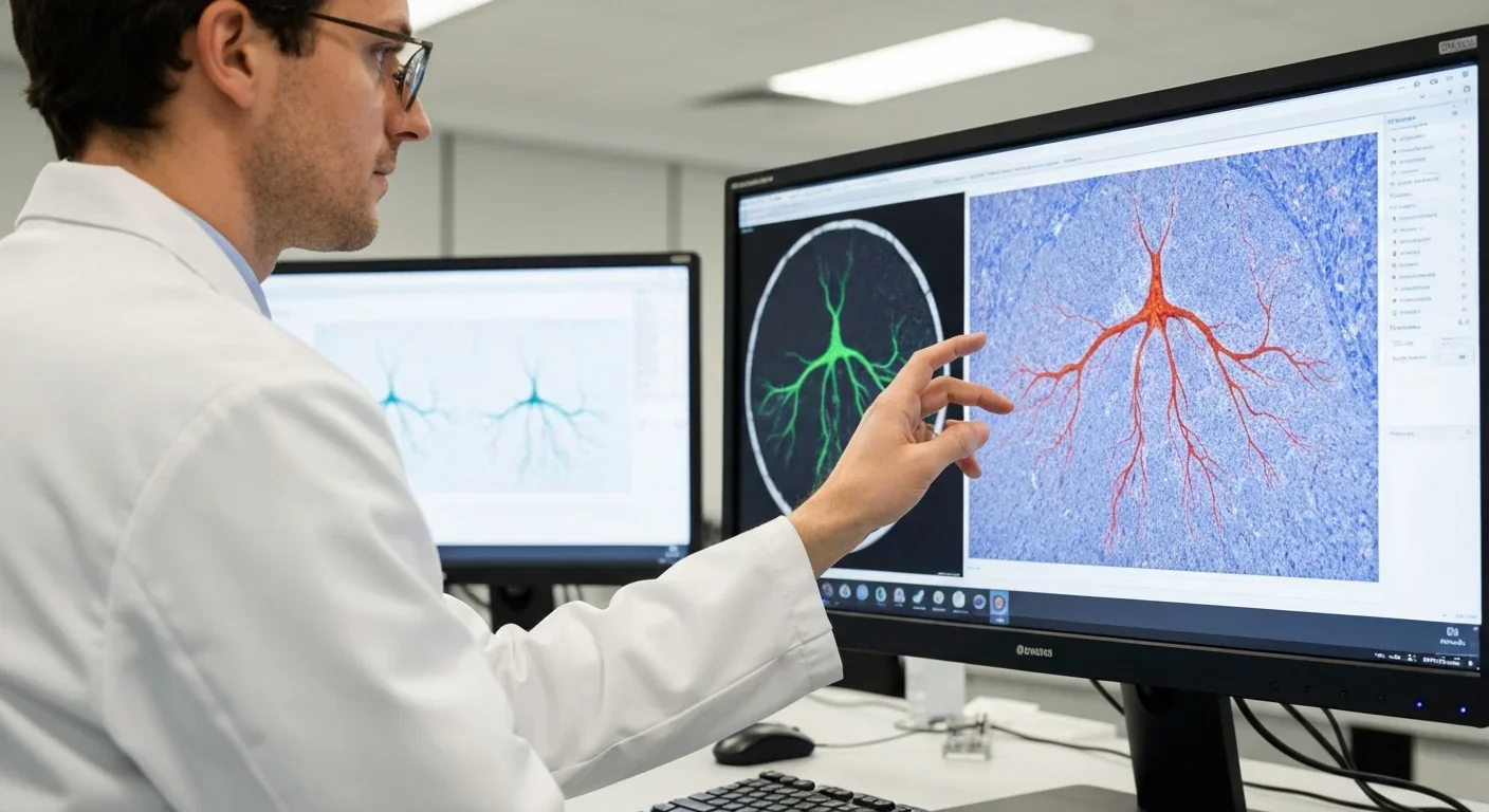
Anatomical Mapping at Unprecedented Resolution. The NIH's SPARC program has invested $15.75 million in a project led by Case Western Reserve and Duke universities to image 100 human vagus nerves using MRI, microCT, and a novel technique called 3D-MUSE. This serial block-face imaging method provides sub-micrometer resolution across the entire nerve, letting researchers trace individual fibers from the brainstem to the abdomen. The resulting three-dimensional scaffold maps, hosted on the SPARC data platform, give any researcher in the world a detailed atlas of where specific organ-bound fascicles sit within the nerve trunk.
Computational Modeling. Those anatomical maps feed directly into computational models that predict which fibers will fire under a given set of stimulation parameters. By simulating different pulse frequencies, amplitudes, and electrode positions on a digital twin of a patient's vagus nerve, engineers can forecast organ-level effects before a single volt is delivered.
The SPARC program's $15.75 million investment aims to image 100 human vagus nerves, creating the first comprehensive atlas that links fascicle position to organ function, a foundation for truly personalized nerve stimulation.
Interferential Current Steering. Perhaps the most dramatic leap comes from a team led by Stavros Zanos at the Feinstein Institutes. Instead of sending a single pulse through the entire nerve, they deliver two slightly offset high-frequency signals, one at 20 kHz and another at 22 kHz, through different electrodes on a multi-contact cuff. These signals interact to produce a localized interference pattern inside the nerve bundle. The result? In pig experiments, they selectively activated lung-related fibers, slowing breathing without triggering throat or voice-box fibers at all.
As neural engineer Kip Ludwig noted, the technique works by making it "a bit harder to activate the stuff you don't want, which is kind of paradigm."
The most commercially advanced example of organ-targeted VNS centers on the spleen. Kevin Tracey's discovery of the inflammatory reflex showed that stimulating specific vagus nerve fibers sends a signal through the splenic nerve, triggering CD4+ T cells to release acetylcholine. That acetylcholine binds to alpha-7 nicotinic receptors on immune cells, suppressing inflammatory cytokines like TNF-alpha and IL-1 beta. It's an elegant biological off-switch for inflammation, and it works without suppressing the entire immune system the way drugs like methotrexate do.
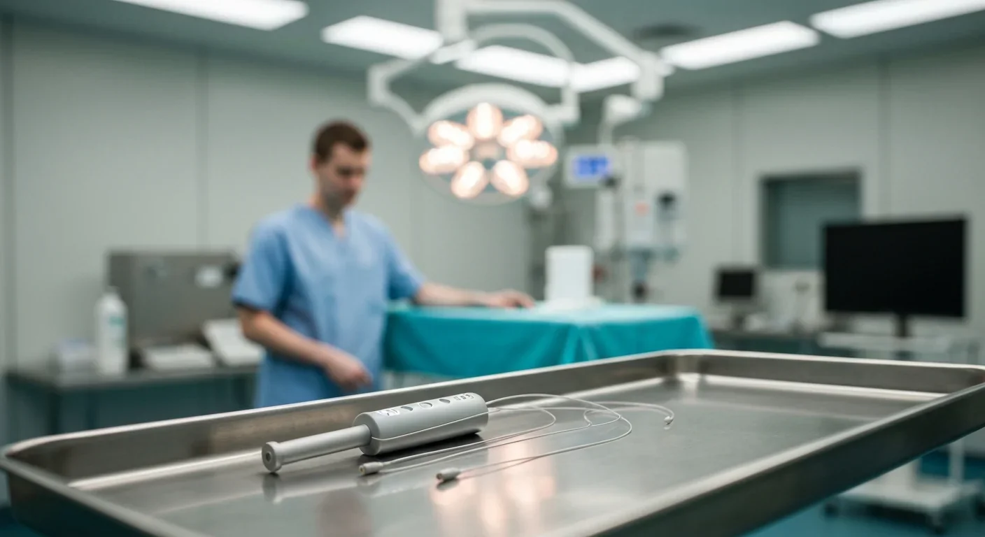
Tracey co-founded SetPoint Medical to translate this into a device. Their system delivers just one minute of daily stimulation through a tiny wireless implant, and the anti-inflammatory effect lasts more than 24 hours. In 2025, it became the first FDA-approved bioelectronic medicine device for rheumatoid arthritis. The company is now investigating the same platform for relapsing-remitting multiple sclerosis.
"This finding provided the conceptual basis for bioelectronic medicine, in which medical devices modulate peripheral nerve activity to treat inflammatory and autoimmune diseases."
- Dr. Kevin Tracey, on the inflammatory reflex discovery
What makes this remarkable isn't just that it works. It's that it works with such precision. SetPoint's researchers spent 20 years identifying exactly which fibers and which stimulation parameters activate the anti-inflammatory pathway without disturbing cardiac or pulmonary function. That's the promise of precision bioelectronic medicine in microcosm.
The clinical applications are expanding rapidly. Heart failure is a major target. Researchers have found that vagus nerve fibers controlling heart rate remain anatomically distinct near the standard cuff site, making cardiac-specific stimulation feasible. A closed-loop VNS system tested in pigs automatically adjusted stimulation based on heart-rate feedback, stabilizing heart rate within 2% to 4% of baseline, essentially acting as an intelligent cardiac buffer.

The gut is another frontier. Early randomized controlled trials of non-invasive vagus nerve stimulation have shown statistically significant improvement in GI symptoms for conditions like IBS and inflammatory bowel disease, with two studies showing reduced TNF-alpha and interleukin-6 in IBD patients. VNS has also been approved for cluster headaches and migraine through the non-invasive gammaCore device.
The perioperative field is watching closely too. A case report documented a 30% reduction in opioid consumption when transcutaneous auricular VNS was applied after surgery, hinting at a future where nerve stimulation replaces some pain medication.
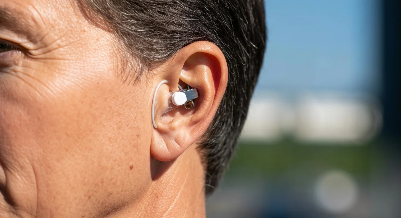
Not all precision VNS requires surgery. The auricular branch of the vagus nerve surfaces at the ear, specifically at the cymba conchae and tragus. Stimulating these spots with small electrodes has been validated through fMRI studies showing activation of the nucleus tractus solitarius and locus coeruleus, key brainstem relays for autonomic control.
A narrative review of 154 studies found that the most common parameters for transcutaneous auricular VNS were 25 Hz frequency, 200 to 300 microsecond pulse duration, and intensity kept below the pain threshold. The safety profile looks reassuring: no serious adverse events were attributed to taVNS across those studies, though 55% of studies didn't formally report adverse events, so some caution is warranted.
Across 154 studies of transcutaneous auricular vagus nerve stimulation, no serious adverse events were reported, though the field still lacks standardized protocols for optimal parameter selection.
Clinical results in post-stroke rehabilitation are particularly encouraging. Multiple RCTs have shown that taVNS at 20 to 30 Hz improves motor recovery, mood, and cognitive function, with benefits persisting up to four weeks after completing treatment. The stimulation also modulates heart rate and heart rate variability, confirming that even non-invasive approaches can produce organ-level effects.
The most exciting frontier may be closed-loop stimulation, where the device monitors physiological signals in real time and adjusts its output accordingly. Think of it as the difference between a thermostat and an open window. A battery-free wireless closed-loop VNS system tested in pigs demonstrated that it could detect rising heart rate and automatically dial down stimulation to prevent bradycardia, all without human intervention.
Combining this adaptive technology with the interferential current steering that Zanos and colleagues developed could create devices that simultaneously target multiple organs while avoiding all off-target effects. Imagine a single implant that calms gut inflammation after meals, stabilizes heart rhythm during exercise, and reduces joint swelling overnight, all by switching between different stimulation programs on the same nerve.
The science is moving fast, but significant hurdles remain. Individual anatomy varies considerably. Fascicle positions differ between the left and right vagus nerves and between patients. The SPARC program's effort to image 100 nerves is designed to capture that variability, but translating population-level maps into personalized stimulation plans requires better intra-operative imaging tools.
Manufacturing precision electrodes at scale is another challenge. A promising low-cost fabrication method using laser-cut silicone and carbon-black composites can produce multi-contact cuff electrodes for about $8.80 per batch of four devices. These electrodes have demonstrated fascicle-selective stimulation in rat sciatic nerves, but scaling from rat to human vagus nerve introduces new complexity.
There's also the standardization problem. Across the taVNS literature, parameter choices vary wildly, frequencies from 1 to 30 Hz, pulse widths from 100 to 500 microseconds, intensities from 0.1 to 60 mA. Without consensus on optimal protocols, comparing results across studies remains difficult.
The trajectory is clear. Just as cardiac pacemakers evolved from crude shockers to sophisticated rhythm managers, vagus nerve stimulation is transforming from a blunt neurological tool into a precision organ-by-organ therapy platform. The anatomical maps are being drawn. The computational models are being validated. The electrode technology is maturing.
Within the next decade, you'll likely see vagus nerve stimulation prescribed the way medications are today, with specific "doses" of electrical frequency, amplitude, and waveform tailored to your individual nerve anatomy and your particular condition. The vagus nerve won't be a mystery wire that doctors zap and hope for the best. It will be a precision instrument, one that lets clinicians send exactly the right message to exactly the right organ, leaving everything else in peace.
For the millions of people living with chronic inflammatory diseases, autoimmune conditions, and neurological disorders that don't respond well to drugs, that future can't arrive fast enough.

Saturn's moon Titan may harbour liquid water beneath its frozen crust, kept from freezing by ammonia acting as a natural antifreeze. New Cassini data suggests the interior could be slush with warm water pockets rather than a global ocean, and NASA's Dragonfly mission launching in 2028 aims to investigate whether this exotic environment could support life.
The cerebellum, long dismissed as merely a motor coordinator, forms dense circuits with the prefrontal cortex that shape cognition and emotion. Disruption of these pathways is now linked to schizophrenia, autism, and ADHD, opening new frontiers in diagnosis and non-invasive brain stimulation therapies.

Research shows the sharing economy often increases total resource consumption through the Jevons paradox and rebound effects. Ride-sharing adds billions of vehicle miles, co-working spaces use more energy per worker, and diffused responsibility erodes conservation behavior. Breaking the paradox requires congestion pricing, accountability design, and matching sharing models to appropriate resource types.

Illusory superiority causes most people to rate themselves above average in driving, intelligence, and ethics. This bias is rooted in metacognitive blind spots, shaped by culture, and carries real costs in healthcare, finance, and leadership. Structured feedback and institutional safeguards can help, but require ongoing effort.

Eastern skunk cabbage generates its own body heat through the alternative oxidase pathway, maintaining temperatures up to 35°C above freezing air and melting surrounding snow. This thermogenic ability, shared by roughly 90 plant species worldwide, reveals a level of metabolic sophistication that challenges assumptions about plant passivity.

America has 28 vacant homes for every homeless person, yet homelessness hit record highs in 2024. Speculative investment, geographic mismatches, and political barriers explain the paradox, while Finland and Vienna show that Housing First and social housing models can work when the political will exists.

Wafer-on-wafer bonding fuses logic and memory silicon at the atomic level, delivering up to 100x interconnect density over traditional packaging. TSMC, Intel, and Samsung are racing to commercialize the technology as AI chips hit the memory bandwidth wall.

Loading featured articles...