Alzheimer's Starts in a Spot You've Never Heard Of
Alzheimer's Starts in a Spot You've Never Heard Of

The Bacterial Sugar Behind Crohn's Disease Flares

How Gut Bacterium Bt Unlocks Fiber's Hidden Nutrition

Metformin Anti-Aging: How a Diabetes Drug May Slow Aging

Precision Vagus Nerve Stimulation: Organ-Targeted

Paraben Science: What Estrogen-Mimicking Preservatives Do
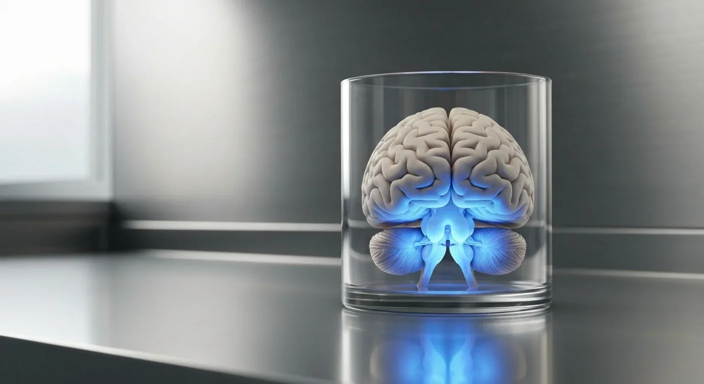
TL;DR: The cerebellum, long dismissed as merely a motor coordinator, forms dense circuits with the prefrontal cortex that shape cognition and emotion. Disruption of these pathways is now linked to schizophrenia, autism, and ADHD, opening new frontiers in diagnosis and non-invasive brain stimulation therapies.
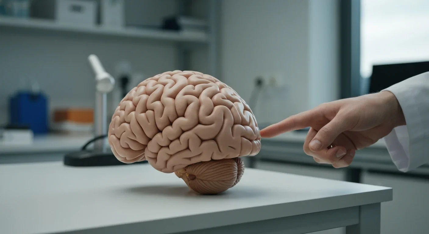
Imagine a brain structure so underestimated that generations of neuroscientists dismissed it as nothing more than a coordination machine. The cerebellum, that fist-sized bulge at the base of your skull, contains more neurons than the rest of the brain combined, roughly 50 to 70 billion of them packed into just 10% of total brain volume. For most of the twentieth century, textbooks described it as the brain's autopilot, fine-tuning your movements so you could walk, type, and catch a ball without conscious effort. That view is collapsing.
A surge of neuroimaging, optogenetic, and clinical research now shows the cerebellum is wired directly into the brain's command center for planning, decision-making, and emotional control: the prefrontal cortex. These dense, bidirectional circuits appear critical for cognitive flexibility, working memory, and emotional regulation. And when they break down, the consequences show up not as clumsy movements but as schizophrenia, autism, ADHD, and depression.
This is the story of how neuroscience's most overlooked structure is becoming its most exciting frontier.
The paradigm shift began slowly and then accelerated. In 1998, Harvard neurologist Jeremy Schmahmann and Janet Sherman published a landmark paper describing patients with cerebellar damage who exhibited a striking pattern of cognitive and emotional deficits: trouble with planning and abstract thinking, spatial disorientation, personality changes, and difficulty regulating emotions. They called it Cerebellar Cognitive Affective Syndrome, or CCAS, and coined the term "dysmetria of thought" to describe how the cerebellum might coordinate thinking the same way it coordinates movement.
For years, skeptics pushed back. But the evidence kept mounting. A 2026 review in PLOS Biology by Diedrichsen and McDougle concluded that "the cerebellum looks like a high-capacity learning engine that can provide a precisely timed predictive signal learned from a very high-dimensional input." Their analysis pulled together decades of electrophysiology, neuroimaging, and clinical data to argue that the cerebellum is not just a motor structure but a universal prediction machine.
The numbers tell a compelling story. The cerebellum's cortical surface area spans roughly 730 square centimeters, far larger than previously estimated, packed into a volume smaller than your fist. This architecture, rich in granule and Purkinje cells organized into thousands of modular units, performs the same computation everywhere. The difference lies in what each module connects to: motor cortex in some regions, and prefrontal, parietal, and limbic areas in others.
"The cerebellum looks like a high-capacity learning engine that can provide a precisely timed predictive signal learned from a very high-dimensional input."
- Diedrichsen & McDougle, PLOS Biology 2026
The cerebellum's reputation as a purely motor structure has roots stretching back to the early 1800s. French physiologist Marie-Jean-Pierre Flourens performed some of the first lesion experiments on animals and observed that removing the cerebellum produced dramatic coordination problems but left "intelligence" seemingly intact. This finding cemented an assumption that persisted for nearly two centuries.
The assumption wasn't unreasonable. Early lesion studies in humans told a similar story: patients with cerebellar strokes presented with ataxia, tremors, and slurred speech. Motor symptoms were obvious and measurable. Cognitive and emotional changes, when they occurred, were subtler and easily attributed to other brain regions or to the general stress of illness.
The first serious cracks appeared in the 1980s. Henrietta and Alan Leiner, along with Robert Dow, published a series of papers arguing that the dramatic evolutionary expansion of the cerebellar hemispheres, particularly a region called the neocerebellum, paralleled the expansion of the prefrontal cortex in humans. They proposed that these two structures had co-evolved because they worked together. The idea was controversial, and many dismissed it.

Then came neuroimaging. When functional MRI became widely available in the 1990s and 2000s, researchers started noticing something puzzling: the cerebellum lit up during tasks that had nothing to do with movement. Language processing, working memory, mental arithmetic, social cognition, the cerebellum was consistently active, and the regions activated were specifically those connected to the prefrontal cortex rather than motor areas.
Randy Buckner's influential 2013 analysis showed that the posterior cerebellar lobes, lobules VI and VII in particular, map onto the brain's association networks, the same networks involved in executive function, default-mode processing, and social cognition. The neocerebellum wasn't just along for the ride. It was a core node in the brain's highest-level circuits.
The anatomical plumbing connecting these two brain regions is surprisingly intricate. Signals from the prefrontal cortex travel down to the cerebellum through a relay station in the pons, arriving as mossy fiber inputs to the cerebellar cortex. After processing by the cerebellum's dense networks of granule cells and Purkinje cells, the output flows through the deep cerebellar nuclei, particularly the dentate nucleus, up through the thalamus, and back to the prefrontal cortex.
This creates what neuroscientists call a "closed-loop circuit": a continuous conversation between the cerebellum and the cortex. The dentate nucleus itself is divided into two functional zones, a dorsal region that handles motor-related output and a ventral region dedicated to cognitive and associative functions. This internal segregation explains how the same structure can simultaneously coordinate your finger movements while refining your working memory.
The dentate nucleus contains two separate functional zones: one for motor control and one for cognition. This dual architecture explains how a single brain structure can juggle movement and abstract thought at the same time.
A meta-analysis of 42 fMRI studies found that cerebellar Crus I, a specific region within lobule VII, consistently activates alongside prefrontal and parietal cortex during verbal working memory tasks. The activation was robust across different experimental paradigms and age groups, from young adults to people in their seventies.
More recently, optogenetic techniques have enabled researchers to directly stimulate specific cerebellar neurons in animal models while recording activity elsewhere in the brain. When scientists use light-sensitive proteins to activate cerebellar output neurons, they can observe downstream changes in prefrontal cortical activity in real time. This level of causal evidence goes far beyond correlation: it proves the cerebellum actually drives changes in how the prefrontal cortex functions.
Why would the prefrontal cortex need a cerebellar co-processor? The answer lies in prediction. The cerebellum operates by building internal models of what should happen next, whether that's the trajectory of your arm during a throw or the expected outcome of a decision. When reality doesn't match the prediction, the cerebellum generates an error signal that helps the rest of the brain adjust.
This "internal model" framework extends naturally to cognition and emotion. Samuel McDougle, a researcher at Yale, describes the cerebellum as "a generalized prediction machine, helping to link our experiences in both motor and non-motor domains." His lab is using fMRI and brain stimulation to map how these predictive circuits support what he calls "motor working memory," the ability to hold and manipulate movement-related information.
A 2025 study in the Journal of Neuroscience provided some of the strongest evidence yet. Researchers tested patients with cerebellar strokes and healthy volunteers receiving cerebellar TMS on a reinforcement learning task. Both groups showed a dramatic reduction, roughly 70%, in the brain's normal prediction-error response, measured via EEG in the frontal cortex. The cerebellum wasn't just passively along for the ride: it was actively generating the error signals that the prefrontal cortex needs to learn and adapt.
Climbing fiber signals, long thought to carry purely motor error information, have now been shown to encode abstract variables like reward prediction errors and surprise during cognitive tasks. The cerebellum's granule cells transform their inputs into a high-dimensional, sparse code that can represent virtually any type of information, motor, sensory, or cognitive, using the same computational architecture.
If the cerebellum helps coordinate thinking and emotion through its connections with the prefrontal cortex, what happens when those connections fail? Increasingly, the answer points toward some of psychiatry's most challenging conditions.
In schizophrenia, the evidence is striking. A 2026 task-based fMRI study found that cerebellar-cortical connectivity was significantly reduced during working memory tasks in patients, with a large effect size (Cohen's d = -1.547). A composite score based on these connectivity measures could distinguish patients from healthy controls with 89% accuracy. Importantly, this connectivity deficit appeared linked to clinical illness rather than genetic risk: unaffected siblings showed normal cerebellar connectivity.

A systematic review across ten resting-state fMRI studies found consistent cerebellar-thalamic hypoconnectivity in schizophrenia, suggesting that the relay between cerebellum and cortex is specifically disrupted. This pattern emerged as one of the most reliable neuroimaging signatures across cohorts.
In autism spectrum disorder, the picture is equally compelling. A review spanning seven different neuroimaging modalities found that cerebellar abnormalities appeared "numerous times across modalities," with reduced cerebellar-prefrontal functional connectivity consistently reported. A bioRxiv study of 751 individuals aged 1 to 21 found that posterior cerebellar growth trajectories predicted social behavior outcomes better than cortical measurements alone.
Cerebellar connectivity measures can distinguish schizophrenia patients from healthy controls with 89% accuracy, pointing toward practical diagnostic biomarkers that could transform psychiatric assessment.
For ADHD, MRI studies have reported smaller cerebellar volumes, and a frontal-cerebellar "When" circuit has been identified as critical for the timing and planning deficits that characterize the disorder. The default mode network, which the cerebellum helps regulate, stays abnormally active during attention-demanding tasks in ADHD, interfering with focus.
Even everyday emotional regulation appears cerebellar-dependent. A 2026 study found that weaker amygdala-cerebellar connectivity correlated with higher levels of emotional dysregulation and smartphone dependence, highlighting how these circuits operate in daily life, not just in clinical populations.
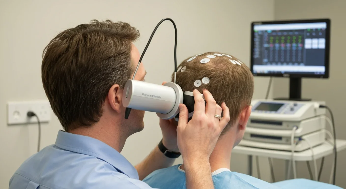
The discovery of cerebellar-prefrontal circuits isn't just rewriting textbooks, it's opening new treatment possibilities. Non-invasive cerebellar stimulation, using techniques like transcranial magnetic stimulation and transcranial direct-current stimulation, can directly modulate these circuits from outside the skull.
A pilot study targeting the right lateral cerebellum in autistic adults showed that a single 20-minute session of theta burst stimulation could significantly modulate connectivity across multiple brain networks, including the default-mode and frontoparietal networks. All participants tolerated the procedure well, with zero adverse events and 100% retention.
"Patients with cerebellar damage sometimes do not demonstrate significant motor deficits, but instead exhibit a range of cognitive symptoms that are robust and replicable."
- Diedrichsen & McDougle, PLOS Biology 2026
For depression, a meta-analysis of tDCS found that 34% of treated patients showed at least 50% symptom reduction compared to 19% on placebo. The UK's National Institute for Health and Care Excellence has confirmed that the approach raises no major safety concerns.
But challenges remain. NICS protocols are not yet standardized, and stimulation fields can inadvertently affect non-target regions. Side effects are generally mild, though rare severe events have been reported. Meanwhile, the CCAS scale provides clinicians with a validated, practical tool for identifying cognitive and emotional symptoms of cerebellar dysfunction, enabling earlier intervention.
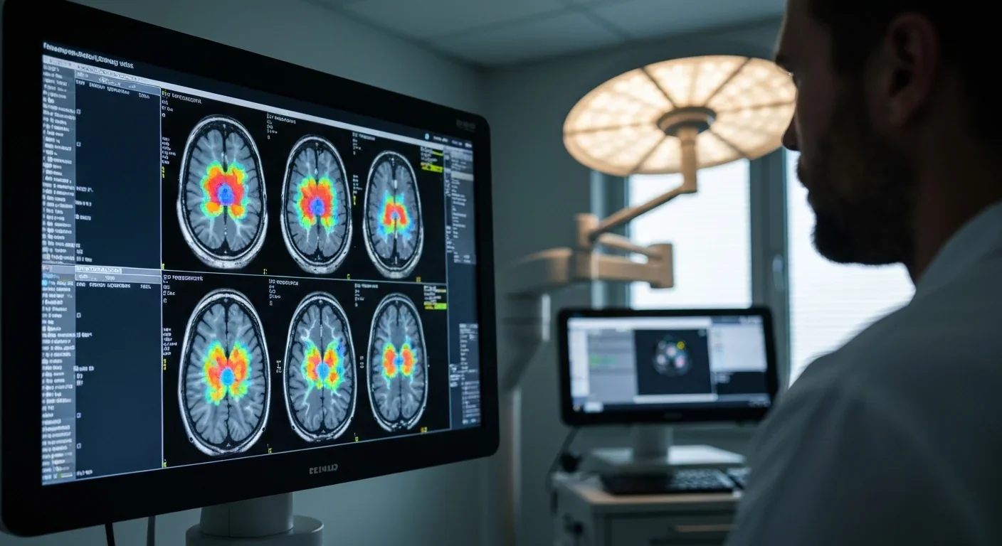
Within the next decade, you'll likely encounter the cerebellum in contexts that would have seemed bizarre just twenty years ago. Psychiatric assessments may routinely include cerebellar connectivity measures, much as blood tests now accompany physical exams. Neuroimaging-based biomarkers are getting close to practical use, with some achieving nearly 90% diagnostic accuracy for schizophrenia.
Research centers across the globe are approaching this differently. In Japan, where the internal-model framework was partly developed, computational models of cerebellar function continue to advance. European centers have pushed forward clinical applications. North American labs focus on cerebellar-targeted therapies using stimulation and pharmacology.
For anyone living with or caring for someone with a psychiatric or neurodevelopmental condition, this research offers a practical message: ask about the cerebellum. The cognitive and emotional symptoms of cerebellar dysfunction are often subtle enough to be missed in routine assessments, and recognizing them opens doors to emerging interventions.
The broader lesson might be about humility. For two centuries, neuroscience was confident it knew what the cerebellum did, and it was wrong. That a structure containing more than half the brain's neurons could be so profoundly misunderstood should make us cautious about what we think we know about the rest of the brain. The cerebellum's quiet revolution is a reminder that the most important discoveries sometimes happen when we finally pay attention to what we've been ignoring all along.

Project Orion was a real 1960s program to reach other stars by detonating 800 nuclear bombs behind a spacecraft. The physics worked and the engineering was feasible, but the 1963 nuclear test ban treaty killed it. It remains the most credible interstellar spacecraft ever designed.
The locus coeruleus, a tiny brainstem structure, degenerates decades before Alzheimer's symptoms appear. Its loss cripples the brain's inflammation control, waste clearance, and sleep regulation. New imaging tools and noradrenergic therapies offer hope for early detection and prevention.

Research shows the sharing economy often increases total resource consumption through the Jevons paradox and rebound effects. Ride-sharing adds billions of vehicle miles, co-working spaces use more energy per worker, and diffused responsibility erodes conservation behavior. Breaking the paradox requires congestion pricing, accountability design, and matching sharing models to appropriate resource types.

Psychological reactance theory explains why banning or restricting things makes people want them more. From Prohibition to the Streisand effect to scarcity marketing, research shows that threatening people's freedom reliably backfires, and autonomy-supportive communication is far more effective.

A carnivorous pitcher plant in Borneo evolved to house bats instead of trapping insects, gaining up to 95% of its nitrogen from bat guano. The plant even built an ultrasonic reflector to help bats find it, revealing that carnivory in plants is a flexible spectrum.
The quantified self movement began as a hacker-ethos pursuit of personal insight, but corporate wearables now funnel biometric data to employers, insurers, and data brokers. With 81% of Americans wrongly believing health apps are HIPAA-protected, a regulatory void enables health data to be sold for pennies while generating anxiety instead of empowerment.

CXL memory pooling lets servers dynamically share DRAM over a cache-coherent interconnect, eliminating the 40% stranded memory waste in data centers. With commercial hardware now shipping and Azure deploying CXL cloud instances, this technology promises to cut memory costs by 50% while enabling composable infrastructure.

Loading featured articles...